
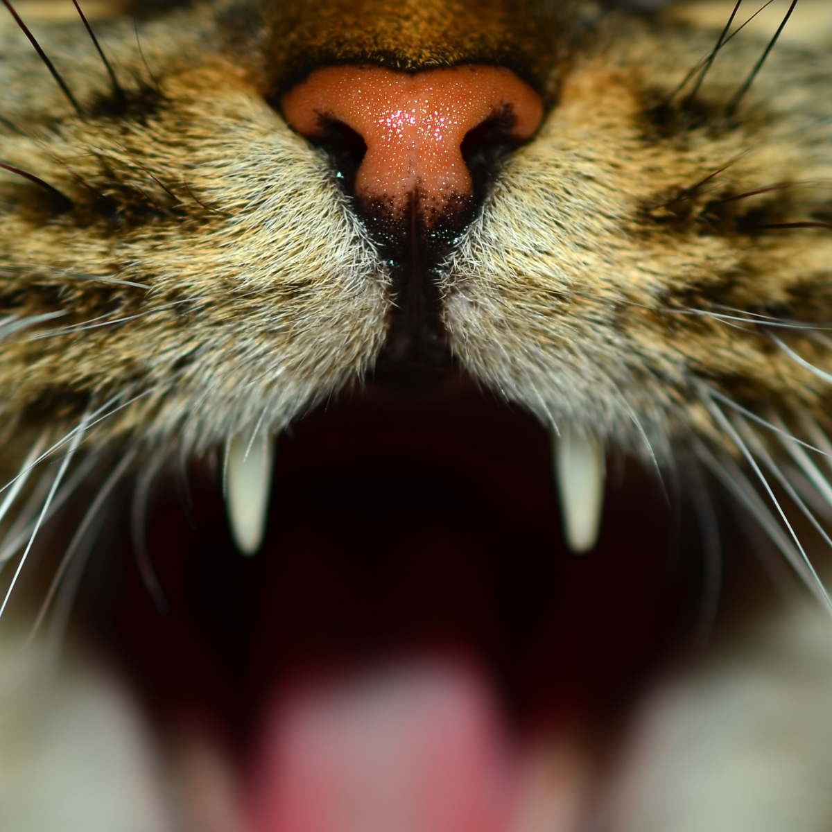
Common Dental Problems
We treat a variety of issues, including:
-
Periodontal disease is the number one health problem in small animal patients. Research suggests that 90% of dogs have some degree of periodontal disease by one year of age. There are generally few or no outward clinical signs of the disease process, and therapy therefore typically comes very late in the disease. This means that periodontal disease may also be the most undertreated disease in our pets.
Unchecked periodontal disease has numerous local as well as systemic consequences. Local consequences include: oronasal fistulas, class II perio-endo lesions, pathologic fractures, ocular problems (including blindness), osteomyelitis, and increased incidence of oral cancer. Systemic diseases which have been linked to periodontal disease include: kidney, liver, respiratory (lung), and cardiac (heart) diseases; osteoporosis, adverse pregnancy effects, and diabetes.
Periodontal disease is generally described in two stages, gingivitis and periodontitis. Gingivitis is the initial stage in which the inflammation is confined to the gingiva. The inflammation is created by plaque. Plaque is made up of bacteria that mixes with glycoproteins and other components of saliva. Gingivitis may be reversed with thorough dental prophylaxis and consistent homecare. Periodontitis is the later stage of the disease process and is defined as an inflammatory disease of the deeper supporting structures of the tooth (periodontal ligament and alveolar bone). As the inflammation invades deeper around the tooth, it results in progressive destruction of the tissues, leading to attachment loss. This can be seen as gingival recession, periodontal pocket formation, or both, and may eventually lead to loss of the affected tooth. Mild to moderate periodontal pocketing may be reduced or eliminated by proper plaque and calculus removal, while more severe pockets require more advanced treatment techniques.
The first clinical sign of gingivitis is bleeding during brushing or probing, or after chewing hard/rough toys. This is followed by redness, swelling, and halitosis (bad breath!). As inflammation worsens and infection reaches deeper parts of the tooth, the bacteria in plaque become more aggressive, worsening the condition.
-
Other than loose teeth, the most common local consequence of periodontitis is an oral-nasal fistula (ONF). ONFs are typically seen in older, small breed dogs, but they can occur in any breed and also in cats. Progression of periodontal disease up the palatal surface of the upper (maxillary) canine tooth is the most common cause of ONF occurance, though it can happen at any upper tooth. The result is a communication between the oral and nasal cavities, resulting in an infection (sinusitis) that may result in significant destruction of nasal structures. Clinical signs include chronic nasal discharge (sometimes with blood and usually seen at the nostril on the same side as the fistula), sneezing, and occasionally anorexia. Definitive diagnosis may require general anesthesia. The diagnosis is made by introducing a probe into the periodontal space on the affected surface of the tooth and root. Interestingly, this condition can occur even when the remainder of the patient’s periodontal tissues are relatively healthy (including other surfaces of the affected tooth). Appropriate treatment of an ONF requires extraction of the tooth and closure of the defect with a mucogingival flap. ONF also frequently occurs after extraction of a maxillary tooth, when a mucogingival flap was inadequate or the pet rubbed or licked sutures out prematurely. If a deep periodontal pocket is discovered prior to development of a fistula, periodontal surgery with guided tissue regeneration can be performed to save the tooth.
-
This occurs when the periodontal loss progresses apically (toward the tip of the root) and gains access to the endodontic system (the blood vessels and nerves inside the tooth, or the pulp), thereby causing infection inside the tooth via bacterial contamination. The endodontic infection subsequently spreads though the tooth via the common pulp chamber and infects the other roots. Extraction is generally the recommended treatment, though sometimes we can salvage a portion of a strategic molar or premolar with a combination of extraction and root canal therapy.
-
These fractures typically occur in the lower jaw or mandible (especially the area of the canines and first molars), as a result of chronic bone loss associated with periodontal disease, which weakens the jaw in affected areas. This condition is most commonly seen in small breed dogs, mostly because their teeth (especially the mandibular first molar) are larger in proportion to their jaws as compared to large breed dogs. Pathologic fractures occur most commonly as a result of mild trauma, or during dental extraction procedures. Some dogs have suffered fractures while simply eating. Although this is typically considered a disease of older patients, cases in dogs less than three years of age have been noted.
Because there is usually a lack of healthy bone remaining, low oxygen tension in the area, these fractures carry a guarded prognosis. There are numerous options for fixation, but the use of wires, pins or plates is generally required. Regardless of the method of fixation, the periodontally diseased root(s) MUST be extracted for healing to occur.
-
Traumatic mandibular fractures are most likely to occur just behind the canine teeth, just in front of or behind the first molars, or very caudally, near the temperomandibular joint.There are a number of different repair techniques such as interosseous wiring, interdental wiring, and splints, all of which utilize the veterinary dental approach of achieving proper, atraumatic occlusion as the first step to fracture healing. It is vital to fully assess each tooth and it's long-term viability. Severely fractured teeth may require extraction or, in the case of strategic teeth, and/or teeth important for anchorage of fracture repair, root canal treatment. Root canal treatment of strategic teeth may be staged for patient comfort, and fracture healing.A second procedure is required in approximately 6-8 weeks after surgical repair is necessary to assess fracture healing, complete any additional dental treatments such as root canal or extractions, and to remove fixation as needed.
-
Fractured or broken teeth can be quite painful! Teeth are covered with a thin layer of enamel - the hardest substance in the body and almost quite literally a rock - which cannot be regrown or regenerated. Once it’s gone, it’s gone. In the middle of a tooth is pulp, which consists of blood vessels and nerves and exits and enters the tooth through the tip of the root, or apex. Between the pulp and the outside of the tooth is dentin. Dentin is also largely inorganic material but it is porous; it is made up of billions of tiny microtubules that radiate out from the pulp like the spokes of a bicycle wheel, and each microtubule is connected to the blood vessels and nerves in the pulp.
When a tooth is chipped and the dentin is exposed (this is also called an uncomplicated crown fracture or UCF), the pulp is indirectly exposed and air, water, food, etc that comes in contact with the dentin can cause pain in the tooth (if you’ve ever had a deep enough cavity and sucked air or cold water over the area, you might know that sharp, painful feeling). Over time, bacteria in the mouth can invade the exposed dentinal tubules and cause an infection inside the tooth, which we generally diagnose with dental x-rays; in one study this was found to happen in almost 1 in 4 teeth with UCF. If no infection is evident, we recommend smoothing the tooth to remove jagged or rough edges, and placing a bonded sealant to block the exposed dentinal tubules and prevent infection.
When a tooth fractures enough that the pulp is exposed (complicated crown fracture or CCF), or if a tooth with UCF shows evidence of endodontic/internal infection, there are only 2 options for that tooth: extraction or root canal therapy.
-
There are several different types of malocclusion in dogs and cats. Some are just bad luck, where one or more teeth are out of normal alignment, and others indicate a genetic issue. The skeletal malocclusions, where the lower jaws are either too long or too short as compared to the upper jaws, are generally genetic and we strongly recommend that affected animals (and their parents) are not used for breeding.
A class 2 malocclusion is one in which the lower jaws are shorter than the upper jaws. This is the most common traumatic malocclusion we see. Because the mandibles are narrower than the upper jaws or maxillas, often the lower canine teeth will impinge on the upper gums, teeth or palate. This condition may be quite painful and lead to other issues, including oronasal fistula, infection, severe wear to other teeth, and even prevent the animal’s jaws from growing to their full potential. Early intervention is vital, and treating the condition in a very young animal might result in a fairly normal adult conformation that doesn’t require further surgery. Selective extractions, orthodontics, vital pulp therapy or root canal therapy are some of the possible treatment options.
A class 3 malocclusion is one in which the lower jaws are longer than the upper jaws. This is a desirable conformation in certain breeds such as Boxers and Bulldogs. While not as often traumatic as a class 2 malocclusion, they can be quite damaging and painful, and selective extractions or odontoplasty (reshaping then sealing a tooth) may be recommended. A very mild class 3 is called an even, or level, bite, and may result in severe wear of the incisors, requiring extraction or root canal therapy.
-
Still a poorly understood disease, tooth resorption occurs in many species, including humans! It is believed to be a derangement of a normal process in which old cells are consumed and new ones laid down. For reasons that are not clear, this process gets out of whack and the tooth structure is eaten away from the outside in, or the inside out. If the resorption is occurring in a portion of the tooth that is exposed to the oral cavity where it may be in contact with saliva, food, water, air, dental instruments, etc., it is quite painful and extraction is recommended. In very specific circumstances, teeth may be treated with crown amputation or even “watchful waiting”. Complete evaluation and imaging under general anesthesia is required to determine viable options for a particular tooth or patient.
-
Canine Chronic Ulcerative Stomatitis (CCUS): Ulcerative lesions on the buccal mucosa where it contacts the teeth is the classic presentation for this condition. The key to keeping this disease under control is strict plaque control, as it appears to be an immune-mediated disease, almost like a contact allergy to plaque. Medical therapy may be considered and consists of a multiple drug and supplement regimen for several months, combined with regular professional cleanings every 3-6 months in addition to strict home care. If/when clinical signs quickly return following regular, frequent dental cleaning, and despite aggressive homecare, it is recommended that we extract any teeth in areas with severe inflammation and start or continue homecare. Full-mouth extractions may be necessary in the future. With full-mouth extractions, patients no longer have a source for plaque to accumulate. This procedure is typically curative and patients show much improvement afterwards. Despite being edentulous, the benefits of decreased inflammation greatly outweigh the lack of teeth, and pets are able to eat and live healthy lives.
Feline Chronic Gingivostomatitis (FCGS) or Feline Caudal Stomatitis: Though our understanding of FCGS has improved in recent years, it is still incomplete. Up to 26% of cats develop FCGS, and cats in multicat living situations are at increased risk. It is believed to be a multifactorial immune mediated disease which causes very painful oral mucosal inflammation. Calicivirus infection is related to development of this disease, though exactly how remains unclear. The disease is characterized by extensive inflammation in the oral cavity - proliferative, ulcerative, or a combination of both - which studies have shown extends beyond the oral cavity and throat to the esophagus and perhaps further into the GI system, even when cats do not exhibit signs of GI disease.
All cats with caudal stomatitis have some level of periodontal disease. FCGS is a very painful condition, and the only oral disease of cats that will stop them eating. It becomes painful to swallow, and many affected cats decrease or stop grooming all together, develop salivary staining on front paws, and drool excessively. There may be foul breath noted. In severe cases, we may recommend placing a feeding tube to administer nutrition and medications prior to and/or after surgery.
Treatment is directed at decreasing the inflammation in the oral cavity, and begins with extraction of all teeth and tooth roots (FME). In select cases in which there is ZERO inflammation around the canines and incisors, these teeth might not be extracted, though extraction at a later time may be necessary if the cat fails to respond to partial mouth extractions (PME).
Almost 70% of cats will respond favorably to extractions, some partially and some completely. There may some continued inflammation in the oral cavity, even if all signs of discomfort have resolved.
We recommend thorough testing prior to surgery so that we can develop appropriate, personalized treatment for your pet before, during and after the procedure. This includes complete blood count, chemistry screen, urinalysis, thyroid, and cardiac screening. In addition, viral testing is recommended: FeLV and FIV should be tested at minimum and calicivirus testing can be submitted if desired. If the proliferative form of the disease is noted, biopsy is strongly recommended to rule out neoplastic (cancerous) changes and will be performed during the extraction procedure.
Long term medical management is associated with poorer outcomes. FIV positive cats are 7 times more likely to develop refractory stomatitis compared to FIV negative cats. If a patient has not responded within 60 days after surgery, further treatment will be recommended. This may include cyclosporine or other immunosuppressive medication. Feline interferon-omega and mesenchymal stem cells (MSC) have also shown promise for treating refractory cases of stomatitis. We expect MSC treatment for FCGS to be commercially available Spring or Summer 2026.
(GSVDS is a clinical study site for Gallant)
-
Though approximately 95% of gingival masses in dogs are considered benign, some can be locally aggressive. Peripheral odontogenic fibroma (POF) is the most common benign mass. Canine acanthomatous ameloblastoma (CAA) is locally very aggressive but does not spread outside the oral cavity. There are many other, malignant oral tumors (melanoma, fibrosarcoma, osteosarcoma etc). We recommend an anesthetized oral exam with dental radiographs, cone beam CT imaging, and a representative biopsy of the mass, and scheduling a second procedure for defiinitive removal and treatment once the mass has been identified, so that appropriate surgical margins may be determined. If imaging reveals no evidence of bony destruction likely secondary to the tumor, debulking with the biopsy and possibly removal of all associated tooth structure may be elected at that time. There is a chance that a second procedure may be necessary, pending results of histopathologic diagnosis, regardless of what the gross and radiographic initial exam indicated.
Presentation and prognosis is different in cats. Oral squamous cell carcinoma (SCC) is the most common oral malignancy in cats, representing approximately 60-80% of oral tumors. Another approximately 10-20% are fibrosarcomas. This means that the majority of oral tumors we find in cats are malignant. The ideal approach for management is early diagnosis through oral examination, radiographic evaluation and biopsy. The next stage in treatment will depend on the results of the biopsy and diagnostic imaging. Oral SCC carries a poor prognosis and therapy options involve radical surgical excision and radiation. Unfortunately, recurrence despite aggressive surgical resection is common. If a more benign tumor is diagnosed, some cats can do well with fairly aggressive surgery as long as the tumor is in the front of the mouth. Tumors far back in the mouth have a much worse prognosis as aggressive surgery is next to impossible given the small jaws and faces of cats as compared to dogs.
-
Enamel hypoplasia and enamel hypocalcification result from disruption of normal enamel development.
Enamel hypocalcification occurs when normal amounts of enamel are produced, but the enamel is incompletely mineralized, making it weaker. The teeth will appear normal after eruption, however they will be more susceptible to fractures. Enamel hypocalcification may also result from trauma to an unerupted tooth, which can affect one or several adjacent teeth, and is the most common acquired cause (often during extraction of a persistent primary tooth).
Enamel hypoplasia occurs when the enamel produced has normal mineral composition, however less enamel is produced than normal. This results in the tooth being incompletely protected with an enamel layer. Enamel hypoplasia may result from a hereditary condition known as amelogenesis imperfecta. This condition causes a decreased amount of enamel matrix to be applied to teeth during development. In these cases, nearly all teeth are affected. A severe systemic infection causing a fever in puppies or nutritional condition may also result in improper enamel production.
It is necessary to protect these teeth to reduce sensitivity and development of periodontal or endodontic disease. Prosthodontic crowns or composite restorations may be recommended for large strategic teeth, which includes canine and carnassial teeth. Bonded sealants are indicated to protect the remaining teeth. This will seal the exposed dentin and help reduce the chance of infection across the dentin, reduce sensitivity, and reduce the adherence of plaque/calculus to the tooth.
-
A persistent primary tooth is a deciduous (baby) tooth that remains when its permanent (adult) counterpart is erupting. A deciduous tooth and its permanent counterpart should never be present at the same time. Consequences of persistent deciduous teeth include increased risk of periodontal disease, malocclusion, and trauma. When a permanent tooth and deciduous tooth are in contact with each other there is loss of normal periodontal tissue and bacteria will accumulate between the crowded teeth. This may lead to permanent damage to and eventual loss of the permanent tooth.
-
Gingival hyperplasia is typically a benign overgrowth of gingival tissue. The cause of this can be genetic, drug-induced, secondary to inflammation from periodontal bacteria, or idiopathic. It is more common in certain breeds such as boxers. Although typically benign, chronic inflammation of this tissue can lead to neoplastic change. If there appears to be an area of excess growth or abnormal tissue, a biopsy sample will be collected and submitted for review to confirm the enlarged tissue is benign. We remove the tissue at the physiological or "normal" gingival margin. The tissue is removed to prevent trauma to this overgrown tissue and to reduce pseudo-pocketing and subsequent periodontal disease. There is a possibility of hemorrhage following surgery, but this should be mild and only last for 24-48 hours as there are no major blood supplies damaged. While it is one of the more painful procedures that we may do, we use aggressive pain management and the gums heal quickly. With young pets with large gingival overgrowth, there is a chance that the excess growth will return to the point that surgery is needed again. However, in older pets and pets with only mild growth, the chance of needing another surgery is low. Aggressive home-care and regular professional dental cleanings will decrease the need for further gingivectomy in the future.
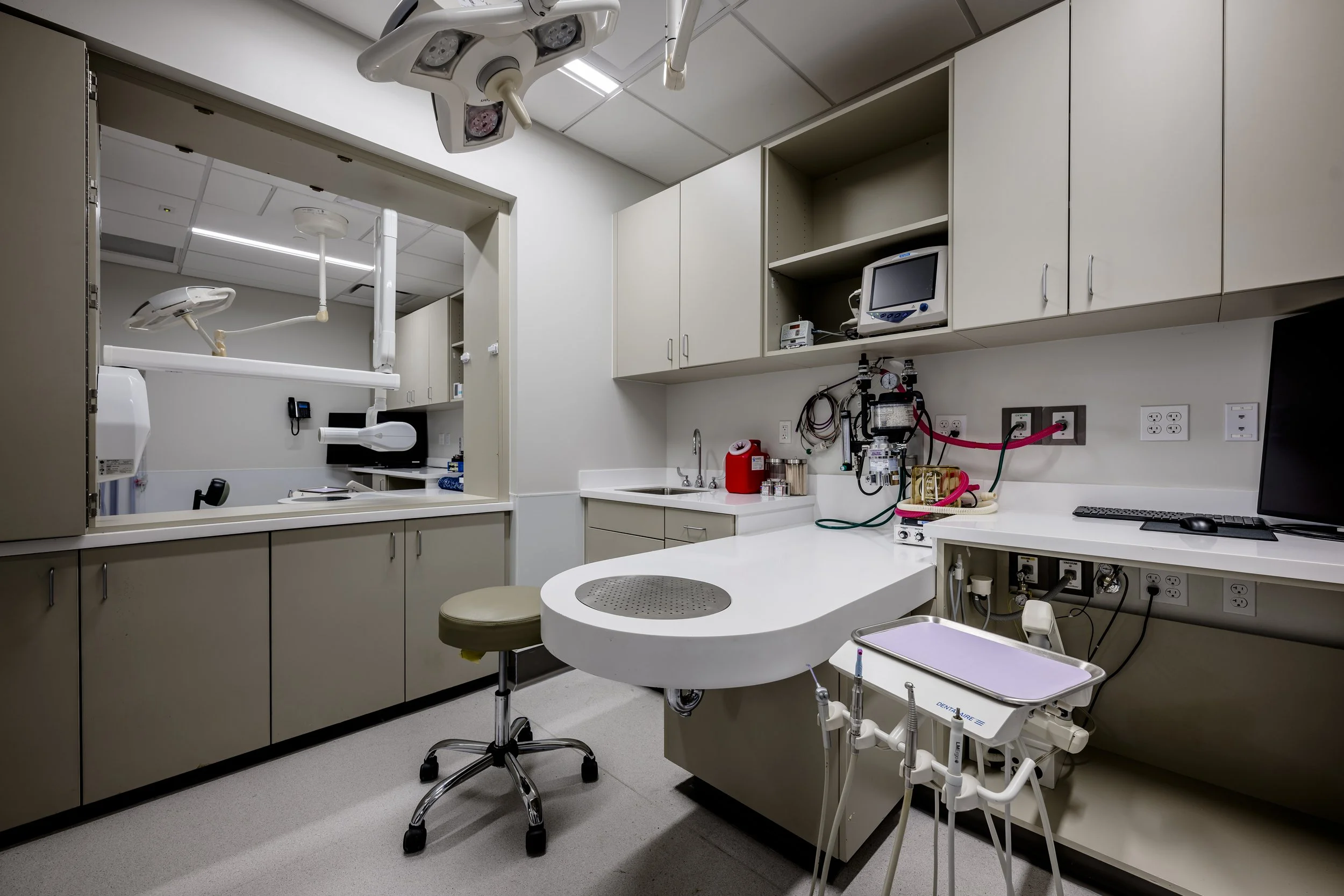
Our Services
-
Extractions
-

Periodontal Therapy
Description goes here -
Fracture Repair
Description goes here -

Endodontics
-
Prosthodontics
Crown Therapy
-

Orthodontics
-
Mass Removal/Oral Surgery
-
Advanced Imaging
-
Intraoral Radiography
-
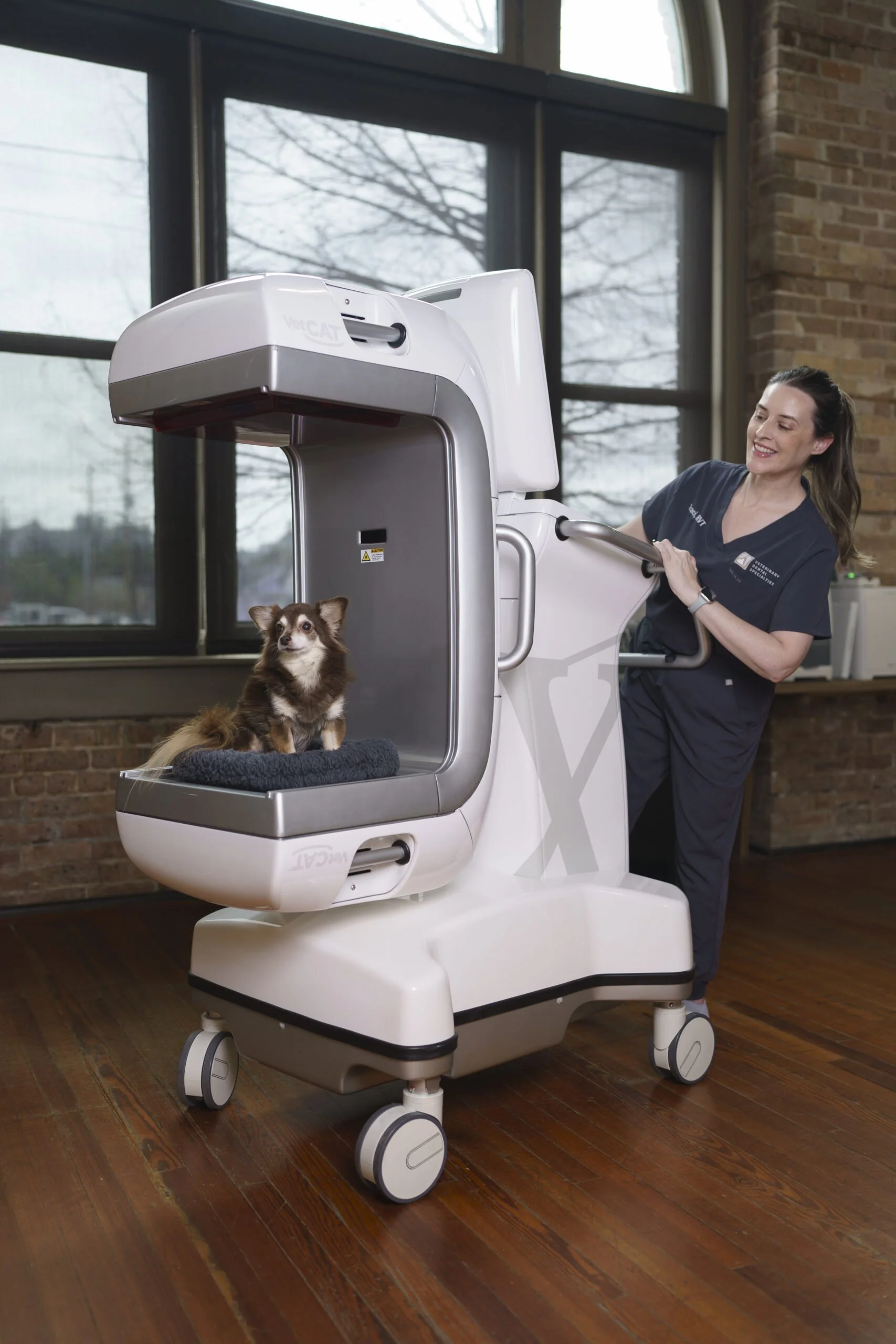
CBCT Bone and Soft Tissue with Contrast
DACVR review available
-

Full Body Radiography
Chest rads, feeding tube placement
Partner Products
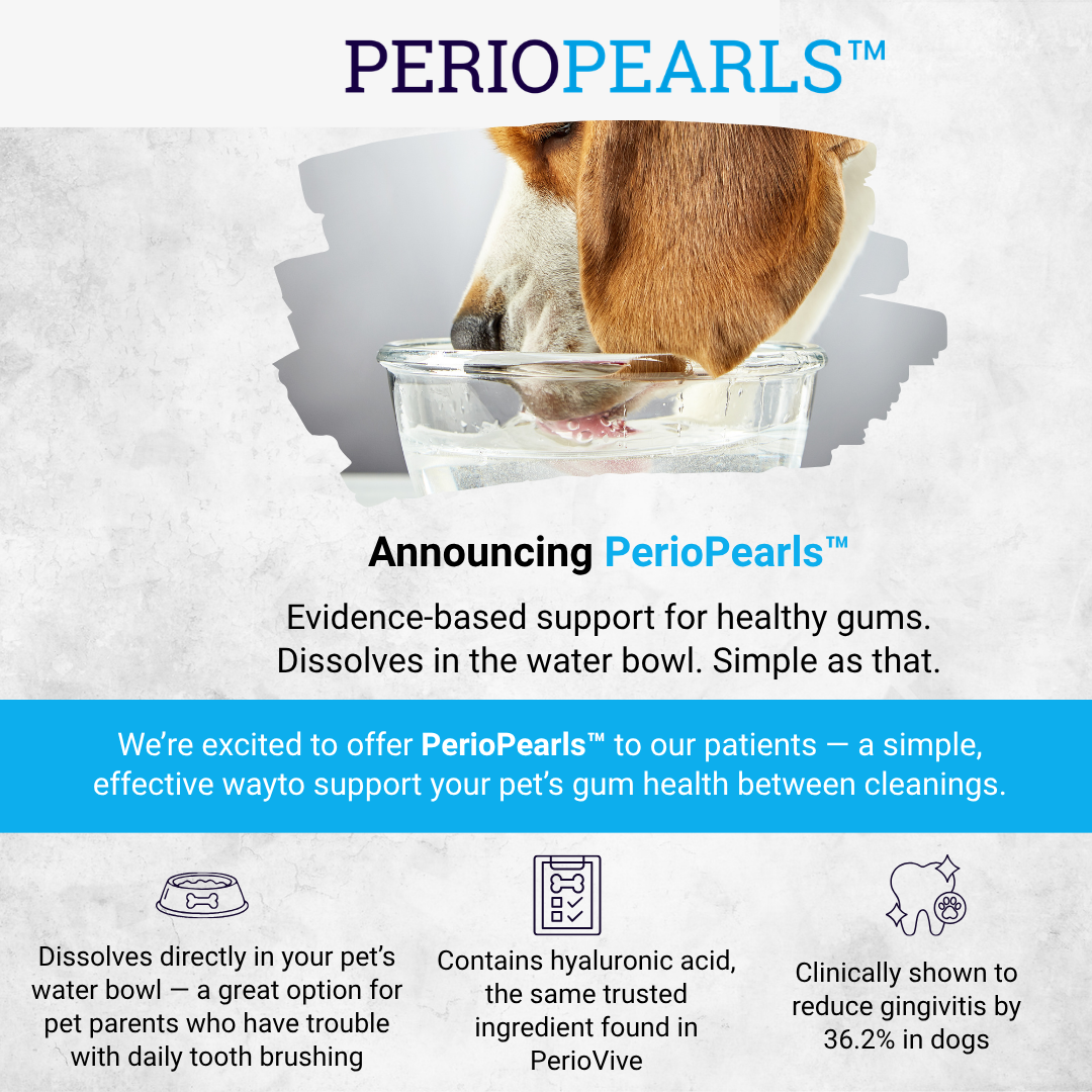
We’re excited to share a new home care option: PerioVive PerioPearls™.
These daily water tablets are designed to support the periodontium, the tissues that surround and support the teeth. Ask our team whether PerioPearls may be a good fit for your pet’s home dental care routine, or go ahead and shop now and enter Gulf South Veterinary Dental Specialties to check out.


Research and Resources
Referring Vet Form
Need to refer a patient to us? Easy, just fill out our form here.
Webinar
Watch Dr. Ribka’s PerioVive webinar on “Periodontal Disease: Treatments, Adjuncts, and an Exciting New Option;”
Enroll your pet in our GingiGuard Oral Gel study
GSVDS is currently involved in a study through July 2026 to slow plaque buildup and bad breath in dogs. If you’d like to participate, review the information below and contact our team to learn more.
Why we are a part of the study: GingiGuard Oral Gel is a promising monthly application of a new technology to slow the recurrence and/or return of plaque, tartar, and bad breath in dogs.
Eligibility: Dogs under 40 pounds with existing tartar and plaque applicable for a dental cleaning.
Owner Requirements: This study requires a dental cleaning and a minimum 4-month follow-up period. The initial oral gel application will be given at the time of the dental cleaning, followed by monthly (in-clinic) exams to check progress and re-application of the oral gel. Study participants are eligible to receive compensation upon completion of the study.
About the Technology: GingiGuard™ Oral Gel, is a topical oral gel designed for monthly application in the management of canine oral health. It provides an innovative surface-acting and localized approach designed to manage plaque, minimize its buildup, and maintain oral health. Unlike other products that require daily effort, GingiGuard Oral Gel is designed for convenient, once-a-month application applied directly to your dog’s gumline.